Happy Clients

Card image cap

Recovery from deteriorating right knee pain
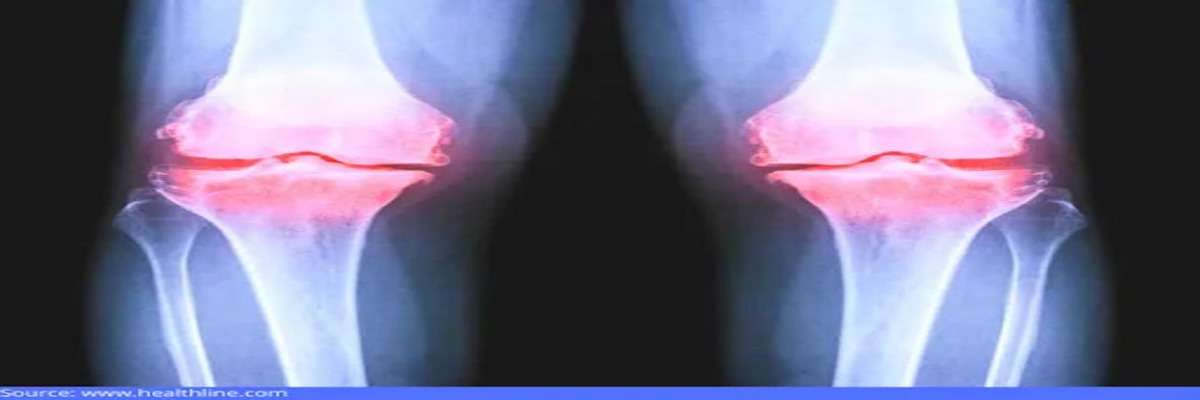
Mrs. Beena Singh came into our office with deteriorating right knee pain. She said her pain started due to a twisting injury while on a short hiking trip with her friends 3 years ago. 

The discomfort was initially relieved after non-surgical treatments like cortisone injections and outpatient PT (physical therapy). However, they only gave temporary comfort, and the pain progressively reappeared a few months later. 

She described her discomfort as a dull ache that is worst on the inside area of the knee. The pain was intensified by activities such as climbing stairs, sitting or getting up, or any activity requiring bending her knees.


TREATMENT PROVIDED TO THE PATIENT


Right Total Knee Arthroplasty

D:\Digicore\UNOSEARCH\Dr. Sandeep Singh\Case Study\knee replacement 2 weeks post Beena Singh\Source After surgery.jpg

Before the surgery, Dr. Sandeep Singh discussed the risks, benefits, and potential complications with the patient and her family.


Several weeks before the procedure, a pretreatment CT scan was taken of the right knee, hip, and ankle. The scan helped in understanding the anatomy and biomechanics of the patient's knee better. The information was used to create a custom implant for the patient. 


During surgery, the highly skilled Dr. Sandeep Singh made an incision to open the front of her damaged joint and removed the injured bone and cartilage. He then replaced them with metal components to recreate the joint surface. After this, he cut the undersurface of the kneecap (patella) and resurfaced it with a plastic button.


Finally, Dr. Singh inserted a plastic medical-grade spacer between the metal replacements to generate a smooth surface.


POST-OPERATIVE ASSESSMENT


Mrs. Singh showed improved movement in her operated knee just after 2 weeks post-surgery. She was relieved that she no longer experienced excruciating pain and was very happy that she could gradually get back to her normal activities.


Card image cap

Bilateral total knee arthroscopy

Mrs. Jayanti Das, aged 71, complained of bilateral knee pain. She first experienced the soreness a few years ago but could not pinpoint when it started. Mrs. Das said the pain started slowly and worsened over time, with a few good days followed by horrible, painful days. The fear of when the pain would recur often kept her stressed out, even on the good days.


She was concerned about the deterioration of her knee discomfort. She was also unhappy about not interacting and socializing with her relatives and kin anymore due to the pain.

Card image cap

Anterior Cruciate Ligament (ACL)

Prateek has been playing football since he was 10. During a practice game about 3 months ago, he felt a popping snap in his left knee when he was moving in for a tackle. His knee started swelling in the next few days but soon began to feel better again. So he went back for practice, that is when his knee just gave in, and he stumbled.


His coach realized that he needed immediate attention and recommended consulting with the renowned Dr. Sandeep Singh.


Prateek underwent physical therapy for a few weeks at CARE Super Specialty Hospital. This was to reduce the knee's swelling and strengthen the muscles around the injured knee. Dr. Sandeep Singh explains that preoperative therapy is essential as people who undergo surgery with a swollen and stiff knee may not be able to regain full ROM (range of motion) after the procedure.


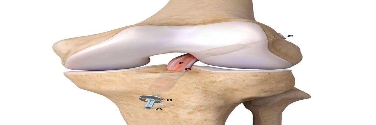
During the ACL reconstruction, the patient was sedated and rendered unconscious. Small incisions were made, one to insert a slender, tube-like camera and the others to provide the accessibility of the surgical tools to the injured area.


Dr. Sandeep Singh removed the injured ligament and replaced it with a healthy segment of a tendon. The graft or replacement tissue was taken from another portion of the patient's knee.


He drilled sockets into the patients' shinbone and thighbone to position the graft accurately and then fastened it to the bones using screws and other devices. The graft served as a support for the growth of new ligament tissue.

Card image cap

Recurring Shoulder Dislocation Fixed with Arthroscopic Bankart Repair
"A dislocated shoulder" – just saying the words is enough to make you feel the pain. Because of all the joints in the body, our shoulders have the highest range of motion. It allows us to lift, grip, and move about effortlessly. When a shoulder is dislocated, it can be tough to function daily. 

Accidents are the most common cause of dislocated shoulders; however, congenital disorders can also cause them.


If your shoulder is dislocated, you will experience severe and instant pain. The apex of the humerus' rotator cuff will feel out of place. Your deltoid muscle will also be unable to move your arm. You may also experience numbness in your arm due to nerve injury.

After your initial shoulder dislocation, you have a 90% chance of relapsing. This is especially true for young people under the age of 20. This is because a dislocated shoulder is more than just a joint problem. It's an injury to the muscle and other soft tissues around it, such as tendons and ligaments. These are the muscles and tissues that aid in the joint's stability. The shoulder joint is compromised when these soft tissues are ripped or injured due to a dislocated shoulder. It's also possible that it will dislocate again.

Some patients' shoulders will dislocate regularly, regardless of their everyday activity. In some cases, involving athletes, mainly contact sports players, surgery to treat a recurring dislocated shoulder may be required. If there is a cartilage tear in the socket of the shoulder joint, this is typically required. This is because the top of the shoulder will need to be sewed back into place for it to be secure again.

Arthroscopic surgery has become a common and highly effective therapeutic option for dislocated shoulders. Because the shoulder joint is placed deep beneath the skin, this is the case. 

Patients with a dislocated shoulder can benefit from the Arthroscopic Bankart Repair treatment. Arthroscopic Bankart repair results are comparable to those of open surgery. With a well-performed arthroscopic treatment, the probability of a redislocation is less than 5%. The goal of surgery is to get individuals back to their normal sporting and work activities.

General anesthesia is used for arthroscopic Bankart repair. It takes around an hour to perform the surgery. Patients are usually admitted to the hospital for one night. After seeing a physiotherapist and meeting with their surgeon, they will be discharged from the hospital the next day.

An Overview of the Case

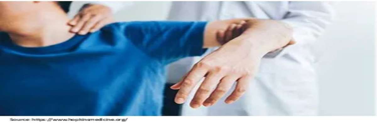
A 20 years young male, Mr. Ajay Bansal, consulted Dr. Sandeep Singh, a seasoned orthopedic doctor in Bhubaneswar, Odisha.  The patient told about a previous shoulder injury that resulted in dislocation. Currently, the patient was in excruciating shoulder pain and had difficulty moving it, with the sensation that it was loose and slipping in and out of the joint.

Dr. Sandeep Singh examined the patient thoroughly and recommended an MRI scan. The MRI examination revealed a dislocated shoulder.

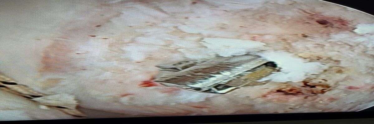
Dr. Sandeep Singh explained the diagnosis and the need for surgery to the patient. Further, Dr. Singh operated on the patient and treated his dislocated shoulder with Arthroscopic Bankart repair.

The doctor prescribed antibiotics and pain medications. Dr. Singh also suggested physiotherapy for a swift recovery. The patient progressively healed and, within a few weeks, regained full range of motion. Rehabilitation enhanced by technology gives near-perfect results post arthroscopic Bankart repair for recurring shoulder dislocation.


Treatment and Prognosis

  • Arthroscopic Bankart Repair


Card image cap

Multi-ligament Knee Injury Treated with Arthroscopic ACL and MCL Reconstruction
Case Presentation

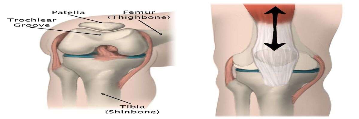
The medial collateral ligament (MCL) and the anterior cruciate ligament (ACL) are two important knee structures. The ACL and MCL, along with other ligaments, work together to keep the knee stable and moving properly.

The ACL limits anterior translation of the tibia onto the femur and contributes to internal and external rotation resistance when the knee is stretched. At 30 degrees of knee flexion, the MCL resists valgus stress. It also aids in reducing anterior and posterior tibia translation and rotation. 

As a result, when both ligaments are damaged, the knee might become unstable in numerous functional planes. Multiple ligament injuries are significantly more complicated than injuries that merely impact the anterior cruciate ligament or the medial collateral ligament.

Treatment for multi-ligament injuries is complex and evolving. The most common treatment for a torn ACL and MCL is surgery to repair the torn ACL, while the MCL injury is treated according to the severity of the injury. The timing of treatment is crucial when both the ACL and the MCL are torn.

Unless the ligament is only slightly ripped, most ligament tears need surgery to repair. Repair surgery, such as knee arthroscopy, may be required if conservative approaches fail. People who participate in sports or have an active lifestyle are more likely to undergo surgery. To discuss all of your choices, you should consult with an experienced orthopedic surgeon in Bhubaneswar, Odisha, like Dr. Sandeep Singh.

An Overview of the Case

Ms. Rinku Biswal, a sportswoman, consulted Dr. Sandeep Singh, one of the best ortho doctors in Bhubaneswar, Odisha. She had difficulty walking and an unstable knee. 

Dr. Sandeep Singh conducted a thorough examination of the patient and recommended an MRI scan. The imaging test revealed MCL and ACL injury.

Dr. Sandeep Singh informed the patient about the injury and the need for MCL and ACL reconstruction surgery. Further, Dr. Singh operated on the patient and performed Arthroscopic ACL and MCL reconstruction. 

The postoperative care included physiotherapy, antibiotics, and pain medications if required. This helped the patient recover quickly. The patient recovered within a few days after surgery and was pain-free and capable of resuming sports. 

After two months of ACL surgery, she won medals for her excellent performance in the field.


Card image cap

Loosened Knee Fixed With Revision Knee Replacement Surgery
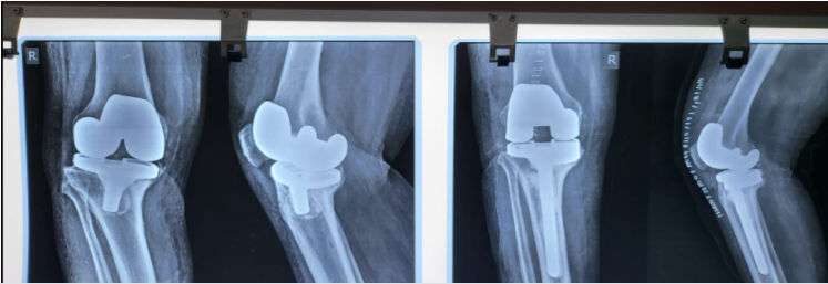
Surgery or a defective implant might cause knee replacement issues. One of the most severe consequences is loosening, and discomfort is the most prevalent. Bone fractures, instability, and major falls can all result from loosening. Almost every severe complication necessitates revision surgery.

Pain, poor joint function, knee instability, and swelling or stiffness in the knee joint are the most typical symptoms of a failed knee implant.

The location of the discomfort can be all over the knee (generalized) or in one specific spot (localized), and it can indicate loosening, wear, or infection. A limp, stiffness, or instability may occur from a loss in knee function. These symptoms and indicators may necessitate revision joint surgery in some patients.

Revision knee surgery using advanced procedures and materials frequently results in significant pain reduction and increased function. Revision knee replacement is a complex procedure that requires an experienced surgeon and proper preoperative preparation to obtain excellent results.

An Overview of the Case

A male patient, Mr. Raghu Nath Panigrahi, consulted Dr. Sandeep Singh, one of the best orthopedic doctors in Bhubaneswar, Odisha. The patient was having difficulty in walking and pain in the right knee. The patient had knee replacement surgery three years ago.

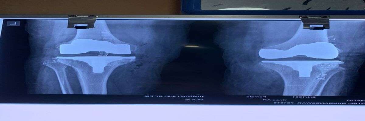
Dr. Sandeep Singh conducted a thorough examination of the patient and recommended an X-ray. The imaging test showed loosening of the knee.

Dr. Sandeep Singh informed the patient about the condition and the need for revision surgery. Further, Dr. Singh operated on the patient and performed revision knee replacement surgery

The patient began walking within a few hours after surgery, and within a few days, he was pain-free and capable of performing routine tasks. The postoperative care included physical therapy, antibiotics, and pain medications if required. 

Treatment and Prognosis

  • Revision knee replacement

Physical Examination & Tests

  • Right knee X-ray


Card image cap

Severe Bilateral Knee Deformity Treated with Knee Replacement

Knee deformity, often known as knock knee, is a condition in which the knees bend inward and "knock" against one another, even when the ankles are apart. It applies too much force to the outside of the knee, causing pain and injury over time. Knock knee is usually bilateral, affecting both legs. 


However, in certain situations, it just involves one knee. Knee deformity is a misalignment of the kneecap that affects individuals of all ages. It can lead to knee discomfort and joint deterioration if left untreated. 


Correcting the deformity will restore knee mechanics and walking ability and lessen pain and slow the progression of a knee injury.


An Overview of the Case

A 60-year-old female patient, Suparna Ghosh, was brought to Care Hospital, one of the top orthopedic centre in Bhubaneswar, Odisha. The patient was having difficulty in walking and severe pain in both knees.


Dr. Sandeep Singh, one of the famous ortho doctor of Bhubaneswar, Odisha, attended the patient. Dr. Sandeep Singh examined the patient and suggested an X-ray, and diagnosed the condition as severe bilateral knee deformity. 


Dr. Sandeep Singh informed the patient and her relative about the condition and the need for immediate surgical treatment. Then, immediately Dr. Singh operated on the patient and performed bilateral knee replacement


After few hours of the surgery, the patient started walking and within several days got complete relief from pain and performed routine tasks efficiently. 


Knee deformity


Card image cap

End Stage Arthritis Treated with Total Knee Replacement Surgery

Arthritis means inflammation of one or more of the joints. It can affect any joint in the body, but it is most common in the knee. The most common symptoms of arthritis are pain, swelling, and stiffness.


Knee arthritis can make it difficult to perform routine tasks like walking or climbing stairs. For many people, it is a major source of lost work time and severe impairment.


End-stage arthritis occurs when the articular cartilage is gradually worn away, resulting in bone-on-bone grinding of the joint surface. End-stage arthritis causes pain as well as a loss of function and movement, significantly limiting daily activities.


In the initial stage, medications and physical therapy can help to alleviate the signs and symptoms. However, when it comes to end-stage arthritis, surgical repair is the only option. 


If arthritic pain is causing disability and nonsurgical treatment is not working, the orthopaedic surgeon may suggest total knee replacement surgery. The surgeon will remove the damaged cartilage and bone and replace them with new metal or plastic joint surfaces to restore knee function.


An Overview of the Case

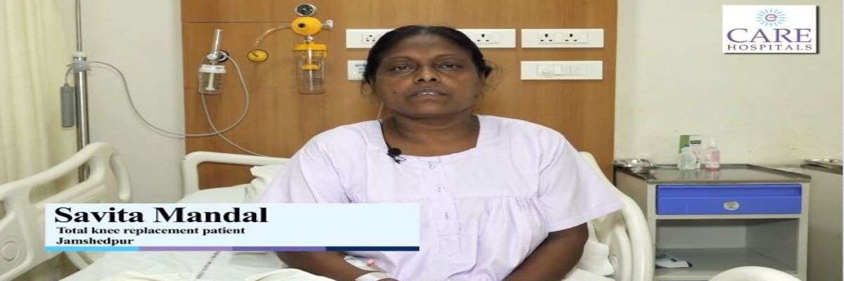
Mrs. Savita Mandal had end-stage arthritis in her right knee, making it difficult to walk and even bend her knee. Under Dr. Sandeep Singh, one of the best bone doctor in Bhubaneswar and team, she underwent a total knee replacement surgery. Within four hours after the surgery, she resumed walking normally and continued living a pain-free life.


Card image cap

Immediate Repair of Right Leg Fracture Resulting from Road Accident

A leg fracture occurs when one of the leg's bones breaks or cracks. Fracture treatment is determined by the location and degree of the injury. A severely fractured leg may necessitate surgery to implant devices into the damaged bone to keep it aligned while it heals. A broken leg must be diagnosed and treated as soon as possible to recover fully.


Delays in diagnosis and treatment might lead to future complications, such as poor healing. Any leg fracture caused by a high-impact trauma, such as a road accident, should be treated immediately. 


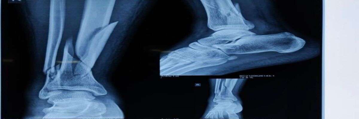
A 20-year-old patient, Mr Rohit Nayak, visited our orthopaedic clinic in Bhubaneswar, Odisha, with trauma in the right leg resulting from a road accident. 


Dr. Sandeep Singh, one of the top orthopaedic surgeon in Bhubaneswar, consulted the patient. Dr Sandeep Singh recommended an X-ray and diagnosed the condition as a right leg fracture. Dr Sandeep immediately operated and fixed the fracture. The next day after the fracture repair, the patient found total comfort and started walking. 


bone-fracture-treatment-in-bhubaneswar
Card image cap

A Case of Severe Sports Injury to Left Knee Treated with Left Lateral Meniscus Repair Surgery

The knee is the body's largest joint made up of the four primary components bones, cartilage, ligaments, and tendons. Because the knee is a complex joint with numerous components, it is susceptible to a wide range of injuries.


Fractures, dislocations, sprains, and ligament tears are among the most frequent knee injuries. In many cases, injuries to the knee involve more than one structure. In sports, often sudden meniscal tears occur. When twisting, cutting, rotating, or being tackled, tears in the meniscus can occur.


In Odisha, a 17-year-young, state-level Kho-Kho player named Santosh Murmu experienced a severe sports injury to the left knee during a tournament. The patient was brought to CARE Super Specialty Hospital, Bhubaneshwar. Dr Sandeep Singh, one of the top orthopedist in Bhubaneswar, handled this case. 


Dr. Sandeep Singh recommended an MRI scan. An MRI showed a meniscus tear in the left knee. Further, Dr. Sandeep Singh performed a left lateral meniscus surgery on the patient. The patient was discharged with full power and control over his limb following comprehensive treatment and guided physiotherapy under Dr. Sandeep Singh's supervision.


Book an Appointment

Enter details,our team would approach to help you as soon as possible.

Phone icon
Call Now